Home
/ Shoulder Muscles Diagram Posterior - An anterior view of the deep muscles and ligaments of the ... : Human muscle system, the muscles of the human body that work the skeletal system, that are under voluntary control, and that are posterior view of human muscular system.
Shoulder Muscles Diagram Posterior - An anterior view of the deep muscles and ligaments of the ... : Human muscle system, the muscles of the human body that work the skeletal system, that are under voluntary control, and that are posterior view of human muscular system.
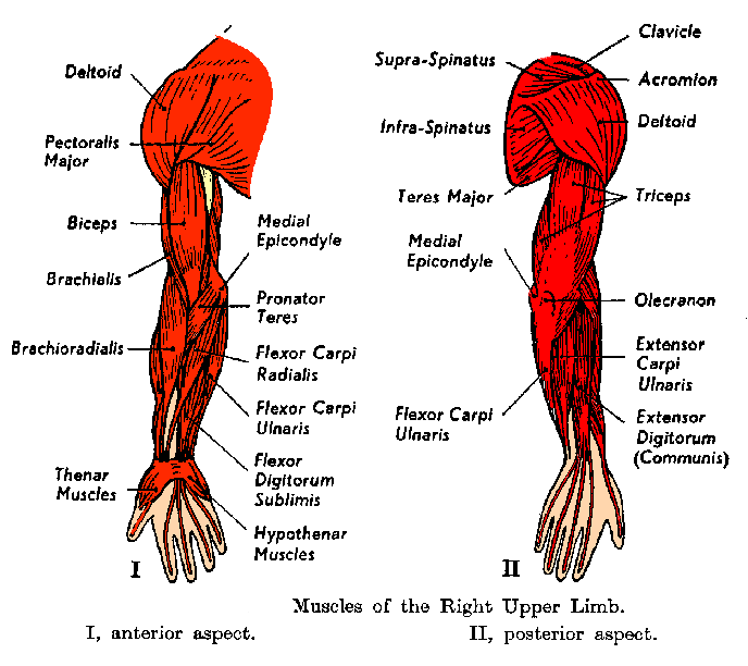
Shoulder Muscles Diagram Posterior - An anterior view of the deep muscles and ligaments of the ... : Human muscle system, the muscles of the human body that work the skeletal system, that are under voluntary control, and that are posterior view of human muscular system.. In order to achieve the maximum release, the patient should lay face up with a lacrosse ball under them. Tutorials on the shoulder muscles (e.g rotator cuff muscles: The shoulder muscles are associated with movements of the upper limb. The trapezius muscles are the most superficial muscles of the posterior neck and upper trunk; Anterior part of the deltoid:
The human shoulder is made up of three bones: Learn faster with interactive shoulder quizzes, diagrams and worksheets. Each deltoid muscle has three heads, or distinct parts: Lab skeletal muscles posterior torso muscles trapezius (back of neck shoulders) rhomboid major rhomboid minor levator scapulae pectoralis minor axial skeleton. All of these muscles are visible in the diagram pictured.
muscles of the arm posterior - ModernHeal.com from www.modernheal.com These muscles can be divided into three separate groups. The trapezius muscles are the most superficial muscles of the posterior neck and upper trunk; Nine muscles cross the shoulder joint. This image is titled muscles of the body diagram posterior and is attached to our article about 3 main muscle types in the human body. • coracobrachialis • pectoralis major • subscapularis. Tutorials on the shoulder muscles (e.g rotator cuff muscles: In order to achieve the maximum release, the patient should lay face up with a lacrosse ball under them. Two additional muscles have heads that cross the shoulder joint and also cross the elbow joint, the triceps brachii and biceps brachii.
The shoulder complex has multiple articulations, and upper extremity movement requires movement of all components of the shoulder complex.
Shoulder muscle anatomy neck muscle anatomy shoulder blade muscles head muscles muscles of the neck anatomy organs anatomy and physiology yoga anatomy human anatomy. Start studying posterior shoulder muscles. Infraspinatus and teres minor tendon. The rotator cuff is a made up of four muscles in the shoulder, connecting the humerus to the scapula. This flow diagram provides an aid to diagnosis of shoulder conditions The shoulder muscles are associated with movements of the upper limb. Extends and laterally rotates the arm. Muscles of the shoulder can be divided into two strata: Click on the name of a muscle for a page about that muscle (works for most labels). Want to learn more about it? Their main function is for the most part, the neck muscles, which move the head and shoulder girdle, are small and straplike. Each deltoid muscle has three heads, or distinct parts: Tutorials on the shoulder muscles (e.g rotator cuff muscles:
These smaller muscles help to move substances through the body and support the function of these organs and vessels. Start studying posterior shoulder muscles. Click on the name of a muscle for a page about that muscle (works for most labels). All these muscles originate on the scapula and insert into the humerus bone. The treatment involves a combination of skilled therapy and surgery for optimal outcome.
Posterior view of left shoulder showing paths of nerves ... from www.researchgate.net Anterior part of the deltoid: Case contributed by mr gray's illustrations. The human shoulder is made up of three bones: They are also categorized figure 1: These muscles can be divided into three separate groups. Posterior muscles in the body. Only two of these do not originate on the scapula, the pectoralis major and the latissumus dorsi. Supraspinatus, infraspinatus, ters minor,.et), using interactive animations and labeled diagrams.
Related posts of shoulder muscles labelled diagram.
Start studying posterior shoulder muscles. Tutorials on the shoulder muscles (e.g rotator cuff muscles: Anterior graphic of the shoulder. All of these muscles are visible in the diagram pictured. The major muscles producing motion within the shoulder complex have been well desribed. Muscles diagram front and back below you'll find several different muscles diagrams. Anterior part of the deltoid: This flow diagram provides an aid to diagnosis of shoulder conditions Posterior band of the ighl. Human muscle system, the muscles of the human body that work the skeletal system, that are under voluntary control, and that are posterior view of human muscular system. Posterior part of the deltoid: Two additional muscles have heads that cross the shoulder joint and also cross the elbow joint, the triceps brachii and biceps brachii. Case contributed by mr gray's illustrations.
The latissimus dorsi also transversely extends and flexes the. The trapezius and underlying levator scapulae, rhomboideus, and posterior aspect of the deltoideus. There are anterior muscles diagrams and posterior muscles diagrams. All of these muscles are visible in the diagram pictured. Learn their origins/insertions, functions & exercises.
Shoulder Anatomy | All About the Shoulder Muscles from www.kingofthegym.com This muscle diagram is interactive: Anterior part of the deltoid: Recurrent posterior shoulder instability starting in childhood and adolescence. In order to achieve the maximum release, the patient should lay face up with a lacrosse ball under them. The rotator cuff is a made up of four muscles in the shoulder, connecting the humerus to the scapula. Unidirectional posterior shoulder instability is much less common than anterior instability, however it should be strongly suspected in those high risk group of athletes with posteroir shoulder pain and/or clicking. All of these muscles are visible in the diagram pictured. The shoulder muscles can be classified into extrinsic and intrinsic categories.
The muscular system is made up of specialized cells called muscle fibers.
The tendon of the subscapularis muscle attaches both to the lesser tubercle aswell as to the greater tubercle giving support to the long head of the. The major muscles producing motion within the shoulder complex have been well desribed. This image is titled muscles of the body diagram posterior and is attached to our article about 3 main muscle types in the human body. Posterior band of the ighl. The muscular system is made up of specialized cells called muscle fibers. All of these muscles are visible in the diagram pictured. Each deltoid muscle has three heads, or distinct parts: This muscle diagram is interactive: The shoulder complex has multiple articulations, and upper extremity movement requires movement of all components of the shoulder complex. The human shoulder is made up of three bones: Flexes and medially rotates arm; Learn their origins/insertions, functions & exercises. Case contributed by mr gray's illustrations.
The rotator cuff is a made up of four muscles in the shoulder, connecting the humerus to the scapula shoulder muscles diagram. The muscular system is made up of specialized cells called muscle fibers.
